Downloads
DOI:
https://doi.org/10.47982/cgc.9.473Published
Issue
Section
License
Copyright (c) 2024 Fred Veer, Telesilla Bristogianni

This work is licensed under a Creative Commons Attribution 4.0 International License.
Abstract
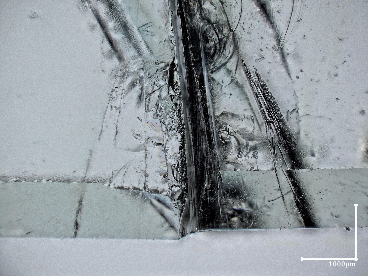
Microscopy has been an indispensable tool for science since the 17th century. It has been used to study glass fractures for decades. The main problem with conventional microscopy is that you can only see a complete image if all aspects of the image are in the same geometrical plane of focus. Digital microscopy is a critical advance since it can combine information in different geometrical planes into a single image. With a transparent material such as glass this opens up new perspectives in that by using the right combination of transmission and reflective lighting both internal defects and complicated 3D surface defects can be imaged. In the field of float glass this allows for the study of edge and surface defects pre-fracture and the damage visible post-fracture. In the field of cast glass this allows for studying the -often complex- network of defects in the bulk. The ability to view and study such internal defects supports research efforts towards the characterization and application of recycled glass. The paper will show the critical aspect of using the right lighting and lenses and the settings needed to obtain high quality images of complex 3D damage. This will be demonstrated based on the microscopic analysis of multiple float and cast glass specimens tested in 4 point bending at the TU Delft Glass lab. Using proper lighting and technique provides a new insight into the fracture processes of glass.